
A new and effective way of completing your root canal therapy utilizes a unique laser system that goes beyond what traditional root canals can offer. It’s called Photon Induced Photo-acoustic Streaming (PIPS), and we are very excited to offer such a high quality service to our patients.
PIPS was invented by a general dentists, Mark Colonna DDS, using the Lares Research PowerLase AT Er:YAG/Nd:YAG laser in one of his operatories about 10:30pm on a Saturday night. This process is so unique that most general dentists and endodontists do not understand the basics of how it works. They have developed tunnel vision thinking that the laser fiber is inserted into the root canal and pulled about burning up anything in its path. This could not be further from the truth. As the name implies, PIPS doesn’t burn up anything, but instead transfers the laser pulse energy into a small controlled explosion of water molecules that sends a shock wave down the root canals to KILL ALL the bacteria and to remove ALL the debris/smear layer inside the root canal’s neuro-vascular system. Yes, I know this statement will disturb many of you in the “Endo World”, but don’t get all bent out of shape until you follow some of the current studies that are going to be published soon.
ACCESS
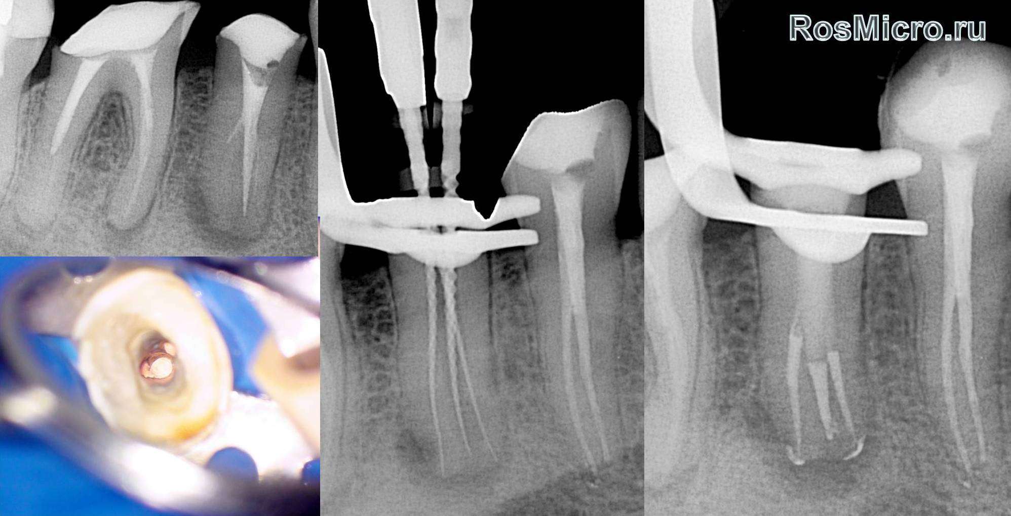
The process of doing a root canal with PIPS starts off just like every other root canal with a well designed access into the pulp chamber so that all the canals are easily instrumented, and of course, a rubber dam.
INSTRUMENTATION
Before putting any instrument into the canals I usually PIPS over the orifice of each canal for at least 20 seconds. I did this on an upper second molar just the other day that I was having a hard time negotiating the canals because of pulp stones. When I was done all the pulp stones were gone and I had a clear pathway into each canal. The Er:YAG laser R12 hand piece was developed to accept different laser tips made of different materials from saphire to quartz. The PIPS tip is a quartz fiber 400? in diameter that has a cone (radial) shaped tip. This cone shape allows the laser pulse to emerge from the end in a radial pattern that sends the acoustic stream of shock waves throughout the entire root structure. This is achieved by placing the Er:YAG PIPS tip into the chamber over each canal sequentially and streaming out the debris from inside the root system for 20 seconds at a time. The dental assistant flows a continuous stream of water into the chamber to allow the laser to explode the water molecules.
Next, File-Ease from Ultradent is introduced to the canals via a Navi-tip. A #6 file is used to find the working length of all the canals using an electric apex locator. The canals are PIPS’d again with a water stream and the process is repeated for the next file sizes up to a#20 or 25 file size. Once this is achieved, the canals are PIPS’d with EDTA then water four times 20 seconds. This process completely sterilizes and removes all the debris and smear layer from the entire root canal system.
OBTURATION
The way we obturate a PIPS endo case is simple. I first use a small paper point to dry up some (but not all) of the water left inside the canals. Using a measured (1 mm short of the apex) #20 Navi-tip from Ultradent, EndoRez (Ultradent) sealer is injected into each canal. This is the cool part. While injecting the sealer into the canals you can see the sealer escape out many accessory canals in the floor of the chamber. This sealer is extremely hydrophilic and it follows water throughout the entire tooth’s neurovascular system. Once the canals are full, a resin coated gutta percha point (Ultradent) is placed into each of the larger instrumented canals (no condensation) and seared off with a touch and heat. Cavit or purple composite flowable resin is then placed to seal off the chamber from oxygen and micro-leakage. Occlusal reduction finishes up the root canal treatment.
Sometimes the EndoRez will “Poof” out the apex, but this is usually OK because the body absorbs it rapidly. As I understand it, this sealer is based on an orthopedic cement used to fixate artificial joints and is very compatible with the bone. I actually have call this a “bone magnet”, but I have been told that is irresponsible and not substantiated in the literature by my Stonehenge “teacher” whom I appreciate more than he knows, so I retract this statement. Notice the MB2 accessory canal going to the pulp chamber. You can actually see these being back-filled as you inject the EndoRez sealer all over the chamber floor.
Why is this technology important?
Dentistry in general has been moving into a new era of minimally invasive treatment. The veneers are getting thinner, cavity preparations are getting smaller, and root canals are also becoming less invasive. As a matter of fact, I will predict that in the future root canals may be done without any instruments being placed inside the the root canal neurovascular system at all. Calcified canals, internal resorption, and extremely curved canal will not pose the problems they currently do today with other systems. The time it takes to do a root canal will shorten and more and more general dentist will find it easier to do endodontics.
Can any dentist with a Er:YAG laser do a PIPS root canal?
No. So far only those dentists with proper training and the Lares Research PowerLase AT, SPA or LightWalker can do a PIPS root canal.
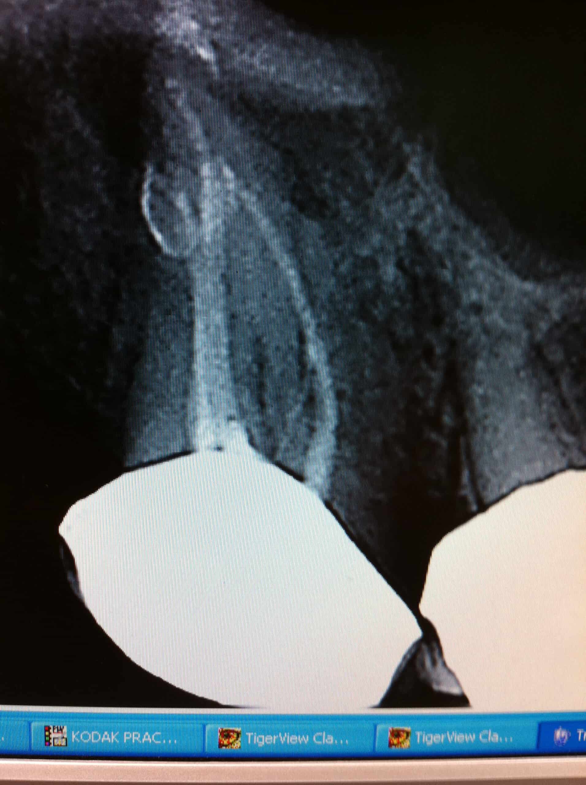
Below are some of our more unusual case which are becoming more usual everyday.
Check out these case and try to count the accessory canals for each root.
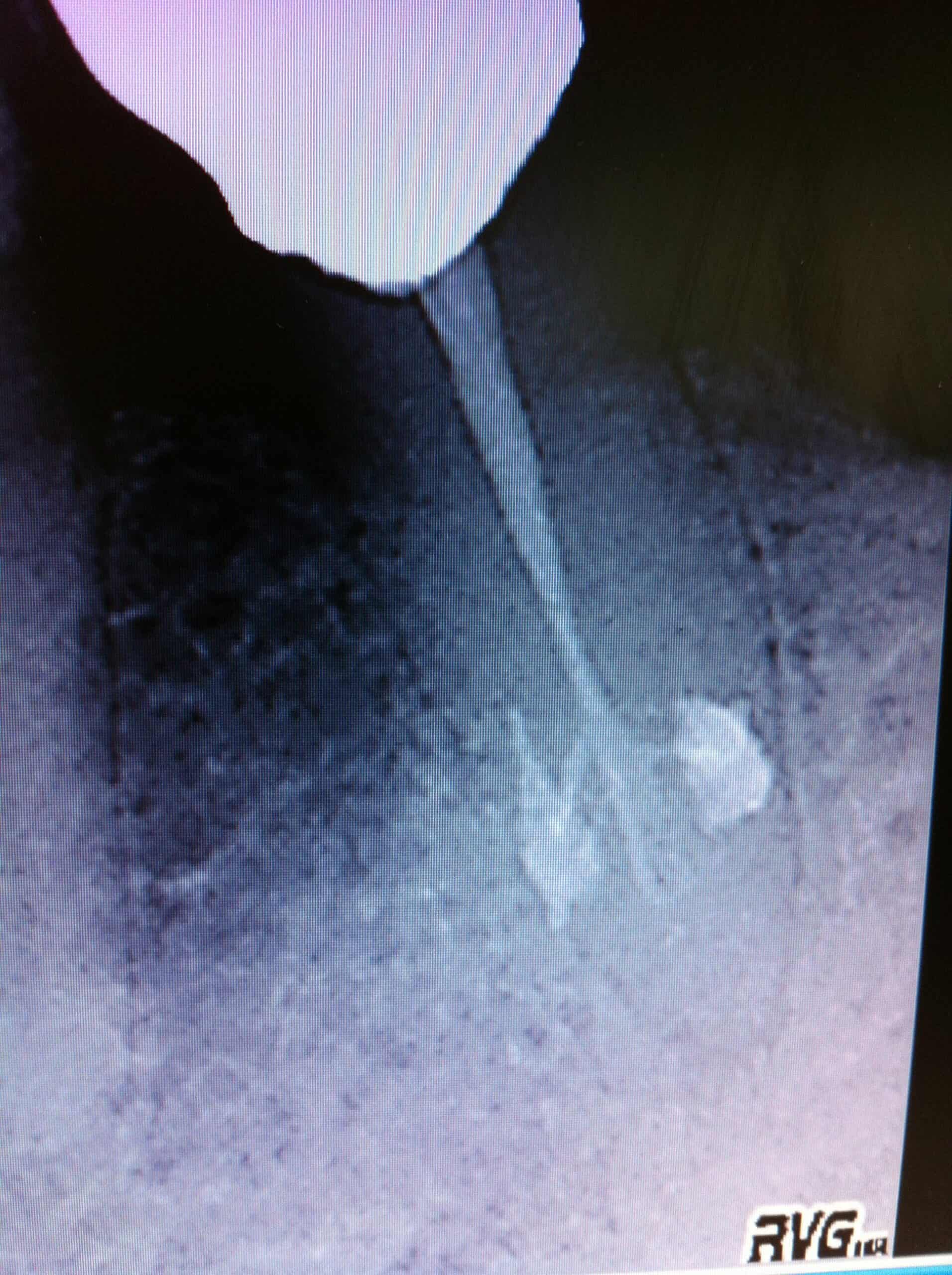
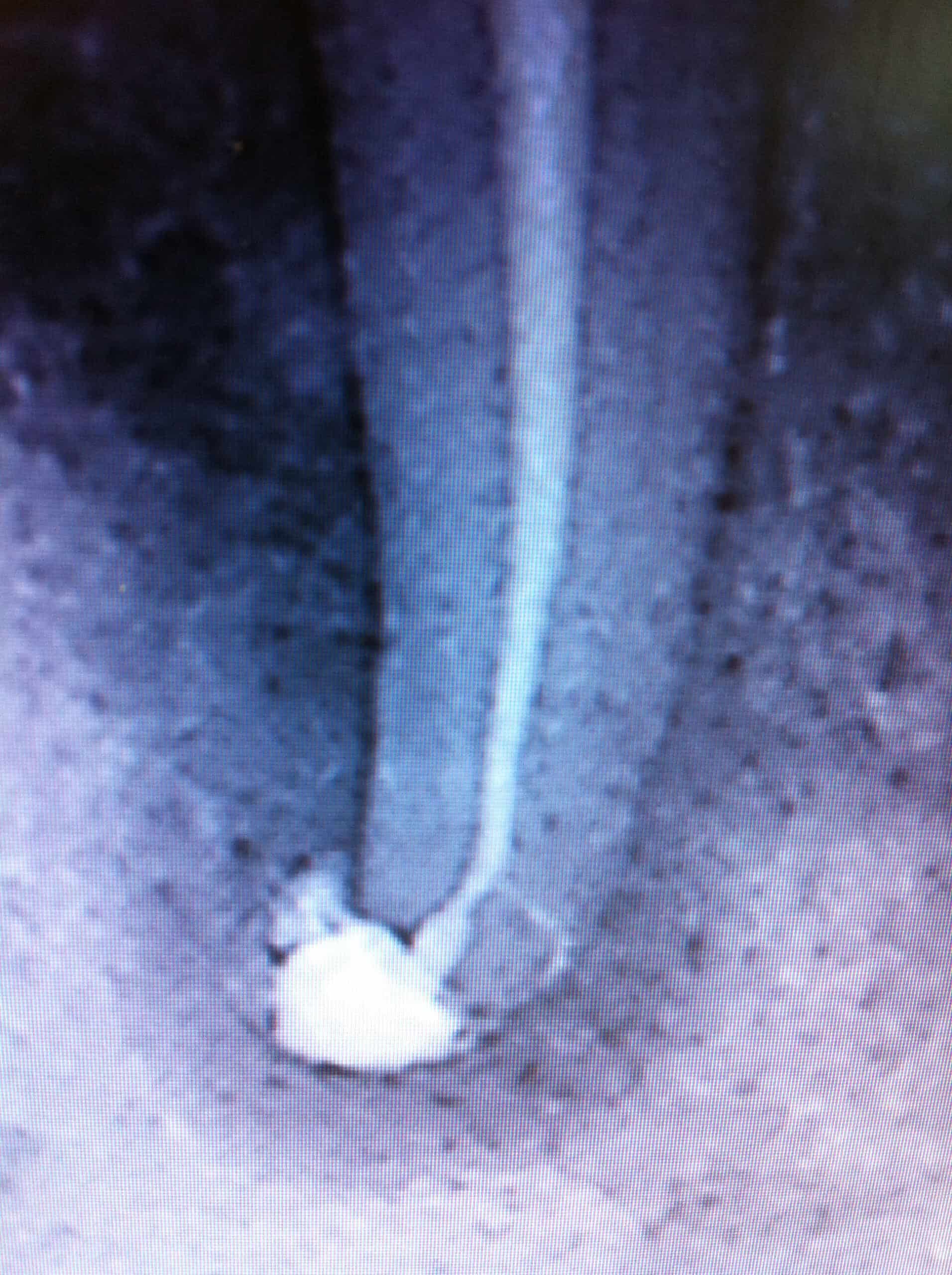
Tough case. Bridge abutment with irreversable pulpitis and calcified canals. This is where PIPS really shines. The canals opened and instrumented to #25 file size.
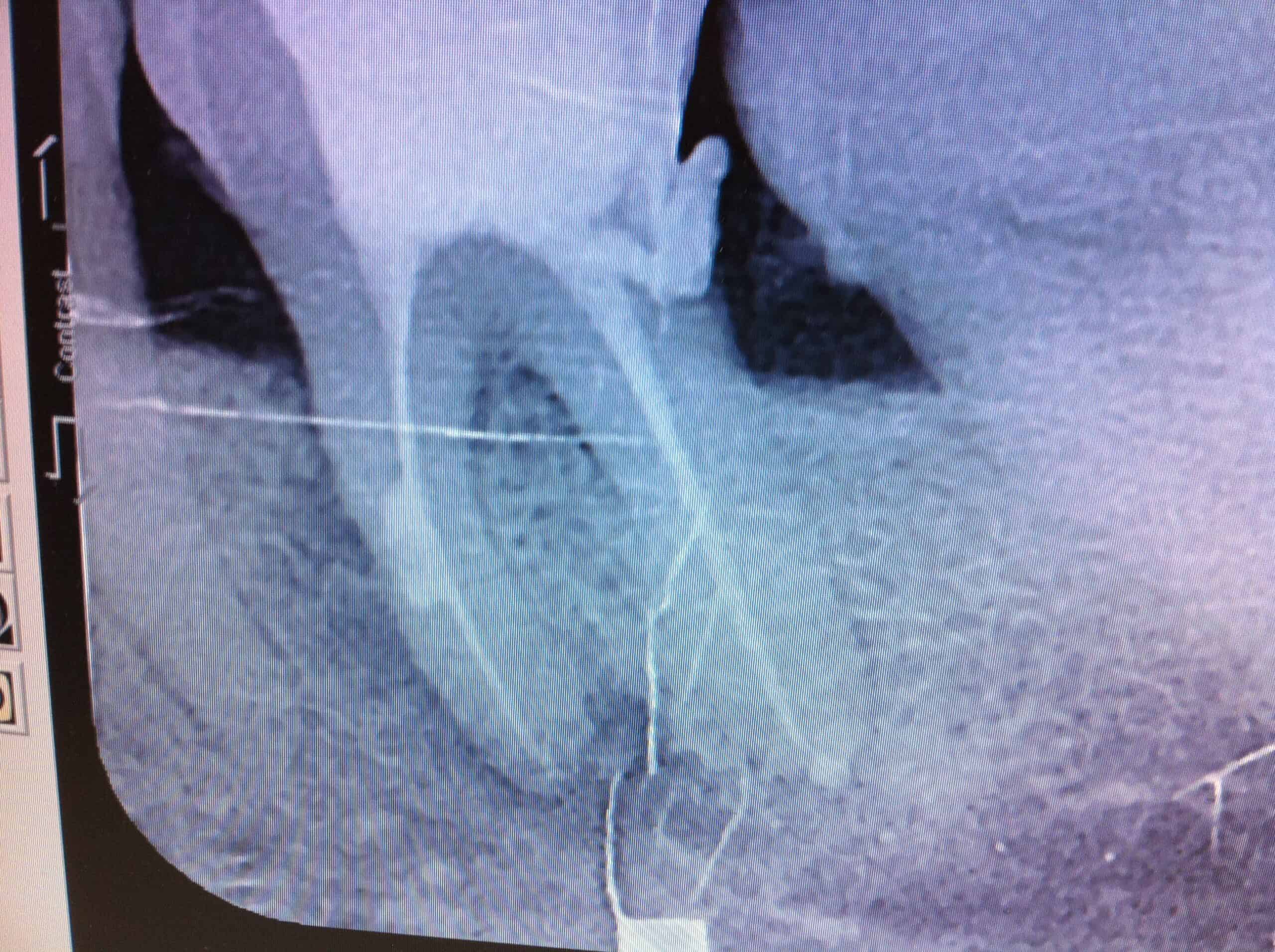
Obturated Internal Resorption
“More information on PIPS Endo with the latest Lares Er:YAG lasers”.
PIPS “Tips”:
This section will be devoted to some of the little problems I’ve noticed during this procedure:
- Before doing any PIPS procedure with your new laser, take a course!!!
- I always use a bite block when using this technique. We were working on #22 when suddenly, without notice the patient bit down. The PIPS tip when down the canal and broke right off! It was wedged in as snug as a bug in a rug. Could not get anything around it. Luckily, the prep wasn’t as wide as the tip all the way down to the apex so I took a long fine pointed gold diamond and chewed it up from the orifice, and then PIPS’d it out.
- Don’t put the tip too deep inside the orifice. I’ve seen that this limits the “PIPS Effect” of rapidly moving fluid in and out of the canal. You can regulate the PIPS effect by moving the tip up and down until you get the churning of the debris out of the canal.
- It is easier to see the PIPS effect if you don’t use a heavy stream of water into the chamber. You need just enough to keep the chamber full and to replace the dirty liquid with clean. This speeds things up by helping you know when you can move to the next step, because your water is clear and no more junk is coming out of the canals.
- Don’t touch the tip of the laser PIPS tip to the inside of the chamber. This will wear the point off rapidly!
- Don’t run the PIPS tip inside the tooth dry. It will not hurt the tooth but it will “abrade” off your PIPS tip quicker with the back splatter.
- Try to open the canals wide enough to get the appropriate NaviTip to within 2mm on the apex. Make sure you test the back pressure needed to express a tiny amount of EndoRez out the tip before placing it into the canal. This will help so you will not shoot a lot of sealer out the apex. Then just barely inject the sealer. Pull the NaviTip back until it is no longer tight in the canal and slowly inject until you see it coming out the orifices.
- The “Perio radial PIPS” tip are thicker and stronger than the Endo PIPS tips and in my opinion work just as well and the endo tips. And they are about $100 less expensive! However, like any other new system, it would be wise to follow the instruction and use the regular PIPS tips for endo, or you will be outside the guidelines set forth by the manufacture, and researchers. They have just come out with a new perio tip that is not as effect as the old because it covers too much of the tip with a sleeving to protect the tip from calculus trauma during WPT. However, I am not so sure the radial tip is that important to the overall PIPS performance because we have done some very good treatments with the point burnt off of our PIPS tips (A good research project for an Endo Grad Resident?)
- Vital or hyperemic pulps are a little harder for the PIPS effect to dislodge than necrotic tissue. Using NaClO will help a lot. Since most of my final endodontic procedures first start off as toothaches, we usually do a pulpectomy first. I do this with files from #6-15, Gates Glidden (2-3), PIPS with peridex (1 minute in each canal or until no more” junk” comes out of the canals), place calcium hydroxide paste (using #15 NaviTip), cotton, IRM, occlusal reduction. This makes the pulp tissue very easy to remove with PIPS/EDTA during the final endodontic procedure.
- If while injecting the EndoRez you see sealer coming out of many other instrumented orifices), don’t assume you need not inject into them independently. There maybe anastomosis that need to be seal through each and every instrumented canal that would be missed otherwise.
- No one is telling you you cannot use your own long tried and true method of doing endodontics with the PIPS technique. You can use it as an adjunct to any existing technique.
- Cleaning the PIPS tip is difficult. We spray it down with viral/bacterial disinfectant and then let it sit wet for several minutes (until the next procedure). However, this maybe a good ways to prevent cross contamination, it is not always the best for your laser. Pay very close attention to how wet with disinfectant the base of the laser tip that is inserted into the R-14 handpiece. If it is wet AT ALL (even damp), then when you fire the Er:YAG it will hit the moisture and splatter it back onto the handpeice’s mirror. After a short while you will notice the laser being less and less powerful to a point it no longer works. Changing the mirror is the only remedy.
- Because the laser arm is balanced to hold its position when you let go of it, it can sometimes cause a problem. I have more than once pushed the arm to the side without paying attention and as I pulled my hand off of the hand piece my glove caught the laser fiber and broke it in half. To avoid doing this you should always act like a robot and watch your hand place the arm/handpiece far enough away from you so this costly mistake doesn’t happen to you too.
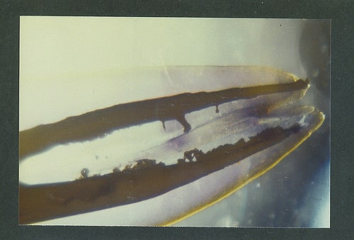
This mirror shows back splatter from a wet PIPS tip.
This mirror was totally burnt out in just 3 months and not allowing the laser to work at all from a wet PIPS tip over time. We have implemented a drying of the laser tip’s base before they are inserted into the R14 handpiece in the hopes of preventing this problem in the future.
Hope this helps! Please feel free to add your own observations in the comment section.
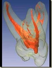
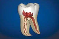
Normal Endodontic Anatomy: Are you doing a good job cleaning it out with your technique? There has got to be a better way…


UPDATE 11/2011
As expected with any new procedure, updates will trickle in from time to time. Recently, my oldest son Dr. Phillip Kraver when to get his certification in both PIPS and WPT. Of course I sat down and picked his brain about what they we’re currently teaching and came to the conclusion that some parts of the procedure had been changed. No game changing information, but different none the less. They now have a Lightwalker Facebook group to discuss all of the procedures so much more information will be able to be found in those discussions: Lightwalker Facebook User Group
- After the initial instrumentation to find the patent canals 5.25% sodium hypochlorite (NaOCl) is PIPS’d into the canals for 20 seconds and then let set through the next size file instrumentation. This is in response to a recent study that will be published that showed 100% killing of E. facalis at the apex of the root canal’s neural vascular system. I previously reserved NaOCl irrigation for canals with hyperemic tissue still present because of its digestive properties. This is because NaOCl interferes with the set of EndoRez sealer that I use in the obturation of the root canal.
- Now they are saying that the PIPS affect is so strong that it is unnecessary to place the PIPS tip directly in or even over each canal. Just placing it into the access and this is apparently enough. This is good news since negotiating the canals with the tip is one of the biggest cause of breakage of the PIPS tips.
References:
1. Journal of Endodontics Volume 26, Issue 8, Pages 454-458, August 2000. Canal Enlargement by Er:YAG Laser Using a Cone-Shaped Irradiation Tip. Shigeru Shoji, DDS, PhD, Hiromi Hariu, DDS, Hiroshi Horiuchi, DDS, DDSc
2. Journal of Endodontics Volume 33, Issue 7, Pages 830-832, July 2007. Evaluation of the Bactericidal Effect of Er,Cr:YSGG, and Nd:YAG Lasers in Experimentally Infected Root Canals. Qian-qian Wang, DDS, Cheng-fei Zhang, DDS, PhD,![]() Xing-zhe Yin, DDS
Xing-zhe Yin, DDS
3. Journal of Endodontics Volume 30, Issue 8, Pages 585-588, August 2004. Q-switched Versus Free-running Er:YAG Laser Efficacy on the Root Canal Walls of Human Teeth: A SEM Study Marouan G. Khabbaz, Mersini I. Makropoulou, Alexandros A. Serafetinides, Dimitris Papadopoulos, Eirini Papagiakoumou
4. Journal of Endodontics Volume 28, Issue 2, Pages 76-78, February 2002. Root Surface Temperature Increase during Er:YAG LaserIrradiation of Root Canals. Yuichi Kimura, DDS, PhD, Kazuo Yonaga, DDS, PhD, Keiko Yokoyama, DDS, PhD, Jun-ichiro Kinoshita, DDS, Yoshiko Ogata, DDS, Koukichi Matsumoto, DDS, PhD
5. Journal of Endodontics Volume 26, Issue 2, Pages 95-99, February 2000. A Histopathological and Immunohistochemical Study of Dental Pulp and Pulpal Nerve Fibers in Rats After the Cavity Preparation Using Er:YAG Laser. Kazunori Takamori, DDS
6. Journal of Endodontics Volume 34, Issue 5, Supplement , Pages e33-e36, May 2008. Lasers in Endodontics: An Online Study Guide
7. Oral Surg Oral Med Oral Pathol Oral Radiol Endod. 2010 Nov;110(5):657-64. Effect of Er,Cr:YSGG laser irradiation on the apical sealing ability of AH Plus/gutta-percha and Hybrid Root Seal/Resilon Combinations. Onay EO, Orucoglu H, Kiremitci A, Korkmaz Y, Berk G. Department of Endodontics, Baskent University School of Dentistry, Ankara, Turkey. eonay@baskent.edu.tr
8. Photomed Laser Surg. 2010 Oct;28 Suppl 2:S63-7. Epub 2010 Sep 23. Apical sealing quality of in vitro apicectomy procedures after using both Er:YAG and Nd:YAG. Leonardi DP, Sivieri-Araujo G, Zielak JC, Baratto-Filho F, Moriyama LT, Berbert FL. Department of Endodontics, Positivo University, Curitiba, PR, Brazil. deleonardi@yahoo.com.br
9. Photomed Laser Surg. 2010 Oct;28 Suppl 2:S43-50. Epub 2010 Sep 23. Investigation of coronal leakage of root fillings after smear-layer removal with EDTA or Nd:YAG lasing through capillary-flow porometry. Michiels R, Vergauwen TE, Mavridou A, Meire M, De Bruyne M, De Moor RJ. Department of Operative Dentistry and Endodontology, Ghent Dental Laser Centre, Dental School, Ghent University, Ghent, Belgium.
10. Photomed Laser Surg. 2010 Oct;28 Suppl 2:S75-8. Epub 2010 Aug 25. Bactericidal effect of Nd:YAG and Er:YAG lasers in experimentally infected curved root canals. Yasuda Y, Kawamorita T, Yamaguchi H, Saito T. Division of Clinical Cariology and Endodontology, Department of Oral Rehabilitation, School of Dentistry, Health Sciences University of Hokkaido, Ishikari-Tobetsu, Hokkaido, Japan. yasuda@hoku-iryo-u.ac.jp
11. Photomed Laser Surg. 2010 Aug;28 Suppl 1:S73-8. Evaluation of antifungal efficacy of erbium, chromium: yttrium-scandium-gallium-garnet laser against Candida albicans. Onay EO, Alikaya C, Seker E. Department of Endodontics, Baskent University, School of Dentistry, Ankara, Turkey. eonay@baskent.edu.tr
12. Photomed Laser Surg. 2010 Aug;28 Suppl 1:S91-6. Effect of Er, Cr: YSGG laser irradiation on Enterococcus faecalis in infected root canals. Yavari HR, Rahimi S, Shahi S, Lotfi M, Barhaghi MH, Fatemi A, Abdolrahimi M. The Department of Endodontics, Tabriz Dental School, Tabriz University of Medical Sciences, Tabriz, Iran.
13. Lasers Surg Med. 2010 Jul;42(5):379-83. Two-dimensional changes and surface characteristics from an erbium laser used for root canal preparation. Roper MJ, White JM, Goodis HE, Gekelman D. Division of Endodontics, Department of Preventive and Restorative Dental Sciences, University of California School of Dentistry, San Francisco, California 94143-0758, USA. mjroperdds@aol.com
14. Lasers Med Sci. 2010 Jun 24. [Epub ahead of print] Histological examination of experimentally infected root canals after preparation by Er:YAG laser irradiation. Kimura Y, Tanabe M, Imai H, Amano Y, Masuda Y, Yamada Y. Division of Endodontics, Department of Conservative Dentistry, Ohu University School of Dentistry, 31-1 Misumido, Tomita-machi, Koriyama City, Fukushima, 963-8611, Japan, y-kimura@den.ohu-u.ac.jp.
15. Lasers Med Sci. 2010 Jun 8. [Epub ahead of print] Influence of the parameters of the Er:YAG laser on the apical sealing of apicectomized teeth. Marques AM, Gerbi ME, Dos Santos JN, Noia MP, Oliveira PC, Junior AB, Zanin FA, Pinheiro AL. Center of Biophotonics, School of Dentistry, Federal University of Bahia, Av. Araújo Pinho, 62, Canela, Salvador, BA, CEP 40110-150, Brazil.
16. Dent Mater J. 2010 May;29(3):246-52. Epub 2010 May 20. Removal of root canal filling materials using Er:YAG laser irradiation. Tachinami H, Katsuumi I. Department of Endodontics and Operative Dentistry, School of Life Dentistry, Nippon Dental University, Tokyo, Japan. hideyuki.t_719@hotmail.co.jp
17. Lasers Med Sci. 2010 Sep;25(5):755-60. Epub 2010 Apr 27. Comparison of dentin root canal permeability and morphology after irradiation with Nd:YAG, Er:YAG, and diode lasers. Esteves-Oliveira M, de Guglielmi CA, Ramalho KM, Arana-Chavez VE, de Eduardo CP. Department for Conservative Dentistry, Periodontology and Preventive Dentistry, RWTH Aachen University, Pauwel Strasse 30, 52074, Aachen, Germany. marcella@usp.br
18. Aust Endod J. 2010 Apr;36(1):6-11. Safety of laser use under the dental microscope. Saegusa H, Watanabe S, Anjo T, Ebihara A, Suda H. Pulp Biology and Endodontics, Graduate School, Tokyo Medical and Dental University, Bunkyo-ku, Tokyo, Japan. h.saegusa.endo@tmd.ac.jp
19. J Oral Sci. 2010;52(1):77-81. Comparison of the effect of Er, Cr-YSGG laser and ultrasonic retrograde root-end cavity preparation on the integrity of root apices. Rahimi S, Yavari HR, Shahi S, Zand V, Shakoui S, Reyhani MF, Pirzadeh A. Department of Endodontics, Tabriz Dental School, Tabriz University (Medical Sciences), Iran.
20. J Endod. 2010 Apr;36(4):725-8. Epub 2010 Feb 6. Comparison of bacterial reduction in straight and curved canals using erbium, chromium:yttrium-scandium-gallium-garnet laser treatment versus a traditional irrigation technique with sodium hypochlorite. Dewsnup N, Pileggi R, Haddix J, Nair U, Walker C, Varella CH. Department of Endodontics, University of Florida, Gainesville, FL 32610-0436, USA.
21. Oral Surg Oral Med Oral Pathol Oral Radiol Endod. 2010 Apr;109(4):e77-82. Morphological assessment of dentine and cementum following apicectomy with Zekrya burs and Er:YAG laser associated with direct and indirect Nd:YAG laser irradiation. de Moura AA, Moura-Netto C, Barletta FB, Vieira-Júnior ND, Eduardo Cde P. Department of Endodontics, Universidade de São Paulo (USP), São Paulo, Brazil. aammoura@usp.br
22. Photomed Laser Surg. 2010 Apr;28(2):161-5. Thermal effects from modified endodontic laser tips used in the apical third of root canals with erbium-doped yttrium aluminium garnet and erbium, chromium-doped yttrium scandium gallium garnet lasers. George R, Walsh LJ. School of Dentistry, University of Queensland, Brisbane, Australia.
23. J Endod. 2010 Jan;36(1):100-4. The synergistic antimicrobial effect by mechanical agitation and two chlorhexidine preparations on biofilm bacteria. Shen Y, Stojicic S, Qian W, Olsen I, Haapasalo M. Division of Endodontics, Department of Oral Biological and Medical Sciences, University of British Columbia, Vancouver, Canada.
24. Photomed Laser Surg. 2010 Feb;28(1):103-8. Analysis of permeability and morphology of root canal dentin after Er,Cr:YSGG laser irradiation. Silva AC, Guglielmi C, Meneguzzo DT, Aranha AC, Bombana AC, de Paula Eduardo C. Department of Restorative Dentistry, School of Dentistry, University of São Paulo, São Paulo, Brazil.
25. Ned Tijdschr Tandheelkd. 2009 Jul;116(7):355-60. [Laser treatment in root canals. Effective by explosive vapour bubbles]. [Article in Dutch] Blanken JW, Verdaasdonk RM. Universitair Medisch Centrum, Utrecht.
26. J Biomech Eng. 2009 Jul;131(7):071004. Evaluation of effectiveness of Er,Cr:YSGG laser for root canal disinfection: theoretical simulation of temperature elevations in root dentin. Zhu L, Tolba M, Arola D, Salloum M, Meza F. Department of Mechanical Engineering, University of Maryland, Baltimore, MD 21250, USA. zliang@umbc.edu
27. Lasers Surg Med. 2009 Sep;41(7):514-9. Laser induced explosive vapor and cavitation resulting in effective irrigation of the root canal. Part 1: a visualization study. Blanken J, De Moor RJ, Meire M, Verdaasdonk R. Department of Dental Materials Sciences, Academic Centre for Dentistry Amsterdam, University of Amsterdam and VU University Amsterdam, Louwesweg 1, 1066 EA Amsterdam, The Netherlands.
28. Lasers Surg Med. 2009 Sep;41(7):520-3. Laser induced explosive vapor and cavitation resulting in effective irrigation of the root canal. Part 2: evaluation of the efficacy. De Moor RJ, Blanken J, Meire M, Verdaasdonk R. Department of Operative Dentistry and Endodontology, Ghent Dental Laser Center, Dental School, Ghent University, De Pintelaan 185/P8, Ghent B-9000, Belgium. roeland.demoor@ugent.be
29. Dent Today. 2009 Jul;28(7):81-2, 84, 86; quiz 87, 80. Lasers as aids for cleaning, shaping, and obturation of the root canal system. Mohammadi Z. Department of Endodontics, Hamedan University of Medical Sciences, Hamedan, Iran. mohammadi_zahed@yahoo.com
30. Lasers Med Sci. 2010 Nov;25(6):805-10. Epub 2009 Jun 23. Bactericidal activity of erbium, chromium:yttrium-scandium-gallium-garnet laser in root canals. Arnabat J, Escribano C, Fenosa A, Vinuesa T, Gay-Escoda C, Berini L, Viñas M. Department of Pathology and Experimental Therapeutics, University of Barcelona, Feixa Llarga s/n, Hospitalet, Barcelona, Spain.
31. Photomed Laser Surg. 2009 Apr;27(2):351-5. The temperature changes in the pulp chamber during cavity preparation with the Er:YAG laser using a very short pulse. Krmek SJ, Miletic I, Simeon P, Mehici? GP, Ani? I, Radisi? B. School of Dental Medicine, Department of Endodontics and Restorative Dentistry, Veterinary Faculty of the University of Zagreb, Zagreb, Croatia. jukic@sfzg.hr
32. Int Dent J. 2009 Feb;59(1):35-46. Laser applications in endodontics: an update review. Mohammadi Z. Department of Endodontics, Hamedan University of Medical Sciences, Hamedan, Iran. mohammadi_zahed@yahoo.com
33. Am J Dent. 2009 Feb;22(1):14-8. Efficacy of calcium hydroxide, Er:YAG laser or gaseous ozone against Enterococcus faecalis in root canals. Noetzel J, Nonhoff J, Bitter K, Wagner J, Neumann K, Kielbassa AM. Department of Operative Dentistry and Periodontology, University School of Dental Medicine, Charité-Universitätsmedizin Berlin, Assmannshauser Str. 4-6, 14197 Berlin, Germany. joern.noetzel@charite.de
34. Alpha Omegan. 2008 Dec;101(4):195-201. The use of lasers for cleaning and disinfecting of the root canal system. Stabholz A, Sahar-Helft S, Moshonov J. Department of Endodontics, Hebrew University-Hadassah School of Dental Medicine, Jerusalem, Israel.
35. Oral Surg Oral Med Oral Pathol Oral Radiol Endod. 2009 Jan;107(1):e47-55. Possibility of root canal preparation by Er:YAG laser. Inamoto K, Horiba N, Senda S, Naitoh M, Ariji E, Senda A, Nakamura H. Department of Endodontics, School of Dentistry, Aichi Gakuin University, Aichi, Japan. kyon@dpc.agu.ac.jp
36. J Endod. 2008 Dec;34(12):1524-7. Epub 2008 Oct 2. Laser activation of endodontic irrigants with improved conical laser fiber tips for removing smear layer in the apical third of the root canal. George R, Meyers IA, Walsh LJ. School of Dentistry, University of Queensland, Brisbane, QLD, Australia.
37. Lasers Med Sci. 2009 Sep;24(5):697-701. Epub 2008 Aug 12. Vibrations produced during erbium:yttrium-aluminum-garnet laser irradiation. Anic I, Miletic I, Krmek SJ, Borcic J, Pezelj-Ribaric S. Department of Endodontics and Restorative Dentistry, School of Dental Medicine, University of Zagreb, Zagreb, Croatia.
38. J Endod. 2008 Jul;34(7):826-9. Epub 2008 May 22. Effects of Er:YAG laser irradiation on biofilm-forming bacteria associated with endodontic pathogens in vitro. Noiri Y, Katsumoto T, Azakami H, Ebisu S. Department of Restorative Dentistry and Endodontology, Osaka University Graduate School of Dentistry, Osaka, Japan. noiri@dent.osaka-u.ac.jp <noiri@dent.osaka-u.ac.jp>
39. J Endod. 2008 Jun;34(6):706-8. Epub 2008 Apr 11. Apical extrusion of root canal irrigants when using Er:YAG and Er,Cr:YSGG lasers with optical fibers: an in vitro dye study. George R, Walsh LJ. School of Dentistry, The University of Queensland Brisbane, Brisbane, Australia.
40. J Endod. 2008 Apr;34(4):474-7. Impact of Er,Cr:YSGG laser therapy on the cleanliness of the root canal walls of primary teeth. Soares F, Varella CH, Pileggi R, Adewumi A, Guelmann M. Department of Pediatric Dentistry, University of Florida, Gainesville, Florida 32610-0436, USA.
41. Lasers Med Sci. 2009 Jan;24(1):7-12. Epub 2007 Dec 18. In vitro preliminary study to evaluate the capability of Er,Cr:YSGG laser in posterior teeth root-canal preparation with step-back technique. Minas NH, Meister J, Franzen R, Gutknecht N, Lampert F, Mir M. Department of Conservative Dentistry, Periodotology and Preventive Dentistry, RWTH Aachen University, Aachen, Germany. nminas@ukaachen.de
42. Lasers Med Sci. 2009 Jan;24(1):59-65. Epub 2007 Nov 20. The impact of an erbium, chromium:yttrium-scandium-gallium-garnet laser with radial-firing tips on endodontic treatment. Schoop U, Barylyak A, Goharkhay K, Beer F, Wernisch J, Georgopoulos A, Sperr W, Moritz A. Department of Conservative Dentistry, Dental School, Medical University of Vienna, Vienna, Austria. curd.schoop@meduniwien.ac.at
43. Lasers Med Sci. 2008 Oct;23(4):443-50. Epub 2007 Oct 24. Histopathological evaluation of pulpotomy with Er,Cr:YSGG laser vs formocresol. Toomarian L, Fekrazad R, Sharifi D, Baghaei M, Rahimi H, Eslami B. Department of Pediatrics, Shaheed Beheshti School of Dentistry, Tehran, Iran.
44. J Endod. 2007 Sep;33(9):1091-3. Epub 2007 Jul 5. Obturation of root canal system treated by Cr, Er: YSGG laser irradiation. Varella CH, Pileggi R. Department of Endodontics, University of Florida, Gainesville, FL 32610, USA. cvarella@dental.ufl.edu
45. J Endod. 2007 Aug;33(8):966-9. Epub 2007 May 18. Interaction between sodium hypochlorite and chlorhexidine gluconate. Basrani BR, Manek S, Sodhi RN, Fillery E, Manzur A. Department of Endodontics University of Toronto, Toronto, Ontario, Canada. bettina.basrani@dentistry.utoronto.ca
46. Lasers Med Sci. 2008 Jul;23(3):247-52. Epub 2007 Jul 21. Effects of Er:YAG laser and Nd:YAG laser treatment on the root canal dentin of human teeth: a SEM study. Kivanç BH, Ulusoy OI, Görgül G. Department of Operative Dentistry and Endodontics, Faculty of Dentistry, Gazi University, Ankara, Turkey. bagdagul@gazi.edu.tr
47. J Am Dent Assoc. 2007 Jul;138(7):992-1002. The antimicrobial efficacy of the erbium, chromium:yttrium-scandium-gallium-garnet laser with radial emitting tips on root canal dentin walls infected with Enterococcus faecalis. Gordon W, Atabakhsh VA, Meza F, Doms A, Nissan R, Rizoiu I, Stevens RH. Department of Endodontology, Temple University, Philadelphia, USA.
48. J Am Dent Assoc. 2007 Jul;138(7):949-55. The use of the erbium, chromium:yttrium-scandium-gallium-garnet laser in endodontic treatment: the results of an in vitro study. Schoop U, Goharkhay K, Klimscha J, Zagler M, Wernisch J, Georgopoulos A, Sperr W, Moritz A. Department of Conservative Dentistry, Medical University of Vienna, Austria. curd.schoop@meduniwien.ac.at
49. Braz Dent J. 2006;17(4):296-9. Er:Yag laser irradiation of the microbiological apical biofilm. Araki AT, Ibraki Y, Kawakami T, Lage-Marques JL. Department of Endodontics, School of Dentistry, University of São Paulo, Avenida Professor Lineu Prestes 2227, 05508-000 São Paulo, SP, Brazil.
50. Int Endod J. 2007 Feb;40(2):112-9. Bactericidal efficacy of Er,Cr:YSGG laser irradiation against Enterococcus faecalis compared with NaOCl irrigation: an ex vivo pilot study. Eldeniz AU, Ozer F, Hadimli HH, Erganis O. Department of Endodontics, Faculty of Dentistry, University of Selcuk, Konya, Turkey. aunverdi@selcuk.edu.tr
52. J Endod. 2008 Aug;34(8):999-1002. Epub 2008 Jun 20. Evaluation of smear layer: a comparison of automated image analysis versus expert observers. George R, Rutley EB, Walsh LJ. School of Dentistry, University of Queensland, Brisbane, Australia.
53. Journal of Endodontics Volume 37, Issue 11 , Pages 1585-1589, November 2011. Efficacy of Smear Layer Removal at the Root Tip by Using Ethylenediaminetetraacetic Acid and Erbium, Chromium: Yttrium, Scandium, Gallium Garnet Laser. Harry Huiz Peeters, DDS, Ketut Suardita, DDS, PhD, Sp. KG, published online 28 September 2011.
54. Journal of Endodontics Volume 37, Issue 6 , Pages 839-843, June 2011. Visualization of Irrigant Flow and Cavitation Induced by Er:YAG Laser within a Root Canal Model. Himeka Matsumoto, DDSYoshito Yoshimine, PhD, DDS, Akifumi Akamine, PhD, DDS
55. J Endod. 2010 Sep;36(9):1580-3. Efficacy of ultrasonic versus laser activated irrigation to remove artificially placed dentin debris plugs. De Moor RJ, Meire M, Goharkhay K, Moritz A, Vanobbergen J. Department of Operative Dentistry and Endodontology, Dental School, Ghent University, Ghent University Hospital, Ghent, Belgium. Roeland.DeMoor@UGent.be